How endotracheal tubes are used in anesthesia is a practical question about protecting ventilation during procedures. An endotracheal tube passes through the mouth or nose into the trachea, creating a controlled airway for selected patients. It may be considered when general anesthesia, surgery, or a patient’s condition makes airway support appropriate. The decision belongs to qualified anesthesia professionals, not a one-size-fits-all checklist.
Safe use involves preparation, placement, confirmation, and ongoing reassessment. Clinicians review the patient’s airway, medical history, procedure, and available equipment before induction. After placement, they assess tube position using clinical findings and continuous waveform capnography, alongside oxygenation and chest movement. The tube is secured, and its depth, cuff, breathing circuit, and ventilation are checked throughout care. Small changes matter. A displaced tube or rising airway pressure needs prompt attention.
This guide explains common tube types, why clinicians choose them, and how teams reduce risks such as sore throat, dental injury, aspiration, or unintended placement. It also covers monitoring and planned removal after anesthesia. Details vary by patient and setting. Practice should follow current professional guidance and local protocols. Technology may change by 2026, but careful assessment and clear communication remain central. Even a familiar airway can surprise a team; that is worth remembering and reviewing after each case. Not every detail is settled by a device. Clinical judgment still matters.
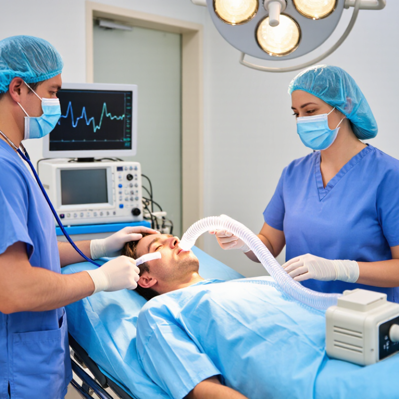
Endotracheal tubes provide a pathway for oxygen and anesthetic gases when a patient needs a secured airway during anesthesia. The tube passes through the mouth or nose into the trachea, helping clinicians support ventilation and reduce the risk of stomach contents entering the lungs. It is one option, not the right choice for every procedure. Patient anatomy, the planned surgery, and health conditions all matter.
A small inflatable cuff near the tube’s tip can help form a seal, while markings on the tube help clinicians assess its position. Placement and ongoing ventilation are checked by trained anesthesia professionals using clinical signs and monitoring equipment, including exhaled carbon dioxide. Details matter. Too much cuff pressure can injure the airway; a tube that shifts can compromise breathing. The team also watches oxygen levels, chest movement, and ventilation throughout anesthesia.
Endotracheal tubes can be especially useful when surgery requires controlled breathing or access to the airway. They may also cause a sore throat, hoarseness, or, less commonly, airway injury. Not every risk is predictable. I think discussions about airway care sometimes sound more certain than real practice allows, because individual responses vary. Patients can ask their anesthesia team why a particular airway device is recommended and what recovery symptoms to report.
Typical Endotracheal Tube Internal Diameter Ranges by Patient Group
Endotracheal tube size is selected according to age, body size, airway anatomy, and the planned procedure. The ranges shown are commonly used starting points and should be adjusted using clinical assessment, ventilation requirements, and available airway equipment. For cuffed tubes, cuff pressure is commonly maintained at approximately 20–30 cmH₂O to reduce the risk of mucosal injury while supporting an effective seal.
Selecting the Appropriate Tube for the Patient and Procedure
Tube selection should match the patient’s airway, surgical position, and ventilation plan. A standard cuffed tube suits many routine adult procedures. Its internal diameter should allow effective ventilation without unnecessary airway trauma. Smaller is not always safer. It can increase resistance and complicate suctioning. For children, sizing should consider age, height, airway anatomy, and local clinical protocols.
The procedure may change the choice. A reinforced tube can resist kinking during prone surgery or head and neck procedures. A preformed oral or nasal tube may improve surgical access.
Lung isolation requires a double-lumen tube or a bronchial blocker, depending on anatomy and clinician expertise. Patients with difficult airways may need a tube that supports guided placement and later exchange.
Before induction, inspect the tube, connector, cuff, and pilot balloon. Confirm that the laryngoscope, suction, and rescue airway equipment are ready. After placement, verify position with continuous waveform capnography and clinical assessment. Secure the tube carefully, especially when the patient will be repositioned. Cuff pressure also needs monitoring. Excess pressure can injure mucosa, while a loose cuff may permit aspiration or air leakage. Tube choice is sometimes uncertain, and experienced clinicians should reassess it when ventilation, surgical access, or patient anatomy changes.
Preparing a patient for endotracheal intubation begins before entering the operating room. Review allergies, fasting status, aspiration risk, dental condition, neck movement, and previous airway records. Ask about loose teeth and removable devices. Difficult airways are uncommon, but consequences can be severe. The UK Fourth National Audit Project reported major airway complications in approximately one of every 22,000 anesthetics. That figure should encourage preparation, not complacency.
Equipment checks must be visible and deliberate. Confirm oxygen supply, suction pressure, masks, laryngoscopes, tube sizes, cuffs, connectors, and waveform capnography. Test the cuff before use. Keep alternative airway devices and emergency access equipment immediately available. The 2022 American Society of Anesthesiologists difficult-airway guidelines support limiting repeated attempts and calling for help early. A clear Plan A, Plan B, and rescue pathway reduces rushed decisions.
Position the patient comfortably, usually with the head and upper body aligned for effective preoxygenation. Explain the procedure in plain language when time allows. Preoxygenate while watching chest movement and oxygen saturation. Capnography should confirm tracheal placement, not auscultation alone. Small oversights still occur, especially during emergencies. I have found that a quiet, repeated equipment check often reveals the missing connector or empty suction canister. Checklists help, but they cannot replace clinical judgment or honest reassessment.
| Stage | Preparation or Action | Key Checks | Purpose and Safety Notes |
|---|---|---|---|
| Patient assessment | Review the medical history, allergies, fasting status, medications, and prior airway or anesthesia records. Assess airway features such as mouth opening, neck movement, dentition, and relevant anatomical findings. | Identify aspiration risk, anticipated difficulty, relevant comorbidities, and the need for additional airway support or a modified plan. | Airway management should be planned by trained anesthesia professionals. The approach depends on the patient, procedure, urgency, and local protocols. |
| Explain and prepare | Confirm patient identity, procedure, consent, and the anesthesia plan. Position the patient appropriately and apply standard monitoring before induction. | Verify functional oxygen saturation, electrocardiography, and blood-pressure monitoring; use additional monitoring when clinically indicated. | Clear communication, adequate monitoring, and an agreed backup plan support safer airway management. |
| Choose the tube | Select an appropriate endotracheal tube type and size based on patient characteristics, clinical indication, and the planned procedure. | Check tube integrity, connector fit, depth markings, and cuff function according to the manufacturer’s instructions and local practice. | There is no single tube size or insertion depth suitable for every patient. Confirm placement clinically and with appropriate monitoring. |
| Prepare airway equipment | Prepare a laryngoscope or video laryngoscope, a suitable tube, a stylet or introducer when indicated, a suction device, a syringe for cuff inflation, and a means of securing the tube. | Confirm that the selected blade and light or screen work, suction is available, and essential equipment is within reach. | Have appropriate rescue devices and a clear escalation plan available, in accordance with the local difficult-airway protocol. |
| Prepare ventilation and oxygen | Check the anesthesia breathing system, oxygen supply, reservoir bag, and ventilation equipment before use. | Confirm circuit connections, gas supply, and the function of relevant alarms and monitors. | Preoxygenation and ventilation decisions should be individualized by the anesthesia clinician and guided by the patient’s condition. |
| Position and preoxygenate | Place the patient in a position suited to their anatomy and clinical needs. Provide preoxygenation when appropriate. | Reassess positioning, mask fit, oxygen delivery, and oxygen-saturation trends. | Positioning and preoxygenation can improve preparation for airway management; use modifications when patient limitations or urgency require them. |
| Insert the tube | After anesthesia induction and any indicated neuromuscular blockade, the trained clinician places the tube using the selected airway technique. | Observe tube passage and patient response; avoid force and follow the planned approach if visualization or passage is difficult. | This is a clinical procedure for qualified professionals. Repeated or forceful attempts can cause harm; follow established attempt limits and escalation guidance. |
| Confirm placement | Confirm tracheal placement using continuous waveform capnography when available, together with clinical assessment. | Assess the capnography waveform, chest movement, breath sounds, oxygenation, and tube depth. Reassess after movement or circuit changes. | Capnography is a key method for confirming and monitoring tracheal tube placement. Clinical signs alone may not reliably exclude esophageal placement. |
| Secure and monitor | Secure the tube, document its position, and continue ventilation and anesthetic monitoring throughout the procedure. | Monitor capnography, oxygen saturation, airway pressures, ventilation, and cuff pressure according to local practice. | Recheck tube position and function if ventilation changes, alarms occur, or the patient is repositioned. |
| Plan removal and recovery | Plan extubation and postoperative airway observation based on the patient’s recovery, airway risk, and procedure. | Confirm readiness for extubation and ensure that appropriate monitoring and rescue support are available. | Extubation is a separate clinical decision requiring trained judgment and a plan for possible airway complications. |
Clinical note: This table is an educational overview, not a procedural protocol. Follow current professional guidance, manufacturer instructions, and local policies; airway management must be performed by appropriately trained clinicians.
Placing an endotracheal tube requires preparation, a clear view, and a calm reassessment. A trained anesthesia professional should confirm the patient, equipment, monitoring, and backup plan before attempting placement. After induction and adequate muscle relaxation, the tube is guided through the vocal cords under direct or video visualization. The cuff should pass just beyond the cords; never force resistance. Small details matter. Note the tube’s depth at the teeth or lips, but treat that number as a reference, not proof of correct placement.
Confirmation starts with continuous waveform capnography. A sustained carbon dioxide waveform over several breaths strongly supports tracheal placement. Check chest movement on both sides and listen for breath sounds across the lungs, while also listening over the stomach. These checks add context, but they do not replace capnography. Reassess oxygenation, ventilation, cuff pressure, and tube security, then document the findings. If the waveform is absent or uncertain, act promptly according to the local airway protocol and clinical situation. Do not wait for a neat explanation. Head movement, transport, or changes in patient position can shift a tube, so repeat checks when circumstances change. Even experienced clinicians can miss subtle signs; a second look is sometimes worthwhile.
Maintaining, Monitoring, and Removing the Tube Safely
A secured tube still needs continuous attention. Check its depth marking after positioning, transfers, and any change in head or neck posture. Watch for condensation, secretions, kinks, and pressure from the breathing circuit. Suction only when indicated, using sterile technique and the appropriate catheter. Small changes matter. A tube that shifts a few centimeters can alter ventilation or become displaced. Confirm ventilation with waveform capnography, chest movement, oxygen saturation, and clinical assessment; no single sign is enough.
Monitoring standards from the American Society of Anesthesiologists call for ongoing evaluation of oxygenation and ventilation during anesthesia, including exhaled carbon dioxide monitoring when an endotracheal tube is used. The UK’s Fourth National Audit Project (NAP4) reviewed 133 major airway complications across anesthesia, emergency, and intensive-care settings. Its findings highlighted that serious airway events can occur beyond initial intubation, including during ongoing care and extubation. That deserves attention. Teams should document tube depth, cuff management, ventilation trends, and any airway concerns, rather than relying on memory during a busy handover.
Plan removal as carefully as placement. Assess wakefulness, breathing, airway protection, secretions, and the risk of swelling or reintubation. Ensure suction, oxygen, and skilled help are immediately available. Remove the tube only when the responsible clinician judges conditions appropriate, then monitor closely for obstruction, stridor, weak ventilation, or falling oxygen levels. A quiet first minute can be misleading. Some patients deteriorate later, and that possibility should shape observation and escalation plans.
It carries oxygen and anesthetic gases into the windpipe during some procedures. It can support controlled breathing.
No. The choice depends on the procedure, airway anatomy, and health conditions. Another airway option may fit better.
They use waveform capnography and assess chest movement, breath sounds, oxygen levels, and ventilation. A carbon dioxide waveform matters.
The small cuff near the tip helps create a seal. Clinicians monitor its pressure; too much may injure airway tissue.
Yes. A reinforced tube may resist kinking when a patient lies face down. Preformed tubes can leave more room near the surgical site.
Breathing may become less effective. Teams recheck its depth and position after movement or repositioning. Small shifts can matter.
A sore throat or hoarseness can occur. Airway injury is less common. Report concerning or persistent symptoms to your care team.
Ask why the device is recommended and which recovery signs to report. The explanation may still feel uncertain; individual responses vary.
This guide explains how endotracheal tubes support safe airway management during anesthesia. It introduces the tube’s purpose, including maintaining airway patency, delivering oxygen and anesthetic gases, and assisting controlled ventilation. It also describes how clinicians select an appropriate tube based on the patient’s age, anatomy, medical condition, and the planned procedure. Before placement, the patient, monitoring devices, ventilation equipment, suction, and airway tools should be prepared, while the airway is assessed and a safe positioning strategy is established.
The article then outlines how endotracheal tubes are inserted by trained professionals and how correct placement is confirmed through clinical assessment and appropriate monitoring. It emphasizes ongoing care, including securing the tube, observing ventilation, checking pressure and position, and responding promptly to complications. Finally, it discusses safe removal after the patient has regained adequate breathing and protective airway reflexes. Overall, understanding How endotracheal tubes are used in anesthesia helps promote organized preparation, careful monitoring, and safer airway management throughout the anesthetic process.

Wigivida Medical